

Safety Training Seminars

The ACLS Cardiac Arrest Algorithm based on AHA 2025 Guidelines delivers a structured, evidence-based approach to adult cardiac arrest management. Review high-quality CPR priorities, shockable and non-shockable rhythms, updated medication timing, airway strategies, and post–cardiac arrest care for improved survival outcomes.
When a patient goes into cardiac arrest, every second counts. Having a clear, systematic plan is crucial for healthcare providers to act quickly and effectively. The Advanced Cardiovascular Life Support (ACLS) Cardiac Arrest Algorithm provides this exact framework. It’s a standardized set of steps designed to maximize the chances of survival from cardiac arrest.
Understanding this protocol helps medical professionals deliver high-quality care during one of the most critical emergencies they will face.
The ACLS Cardiac Arrest Algorithm is a structured guideline developed by the American Heart Association (AHA). It outlines the sequence of actions healthcare providers should take when an adult patient is in cardiac arrest. The main goal is to restore a stable heart rhythm and spontaneous circulation as quickly as possible. The algorithm prioritizes high-quality CPR, early defibrillation, and advanced interventions.
The process is designed to be a continuous loop of assessment and treatment, allowing the response team to adapt to the patient’s changing condition.
The ACLS Cardiac Arrest Algorithm is broken down into a few key actions that are repeated in cycles. Let’s explore each one.
As soon as cardiac arrest is confirmed, the first and most important action is to start high-quality cardiopulmonary resuscitation (CPR). This is the foundation of the entire algorithm.
High-quality CPR involves:
CPR keeps oxygenated blood flowing to the brain and other vital organs until the heart can be restarted.
After two minutes of CPR, the team briefly pauses to check the patient’s heart rhythm using a monitor or defibrillator. The rhythm will determine the next course of action. Heart rhythms are categorized as either “shockable” or “non-shockable.”
If the monitor shows Ventricular Fibrillation (VF) or pulseless Ventricular Tachycardia (pVT), a shock is needed. These are chaotic electrical rhythms that prevent the heart from pumping blood effectively.
If the monitor shows Asystole (a flat line) or Pulseless Electrical Activity (PEA), a shock is not effective. In PEA, there is electrical activity on the monitor, but the patient has no pulse.
Medication is a key part of the ACLS algorithm, especially after the first couple of cycles.
Medications are typically given through an intravenous (IV) or intraosseous (IO) line.
The algorithm is a loop that continues until the patient achieves Return of Spontaneous Circulation (ROSC) or the team decides to stop resuscitation efforts.
A typical cycle looks like this:
During resuscitation, establishing an advanced airway, such as an endotracheal tube or a supraglottic airway, can be beneficial. Once an advanced airway is in place, ventilations can be given every 6 seconds without pausing chest compressions.
Quantitative waveform capnography is used to monitor the amount of carbon dioxide (CO2) in exhaled air. This tool helps confirm airway placement and monitor the effectiveness of CPR. A sudden increase in the capnography reading can be an early sign of ROSC.
Understanding the ACLS Cardiac Arrest Algorithm on paper is one thing, but mastering it in a real-life emergency requires hands-on practice. The ability to perform these steps smoothly and efficiently as part of a team can make a significant difference in patient outcomes.
At Safety Training Seminars, we provide official American Heart Association (AHA) ACLS courses that give you the skills and confidence you need. Our programs are designed for busy medical professionals, with a convenient blended format. You complete the online portion at your own pace and then come to one of our 70+ locations for a short, in-person skills session. You’ll receive your AHA certification card the very same day.
If you’re ready to sharpen your life-saving skills and become an expert in advanced resuscitation, we encourage you to book an ACLS class with us. Join the thousands of healthcare providers who trust Safety Training Seminars for their certification needs.
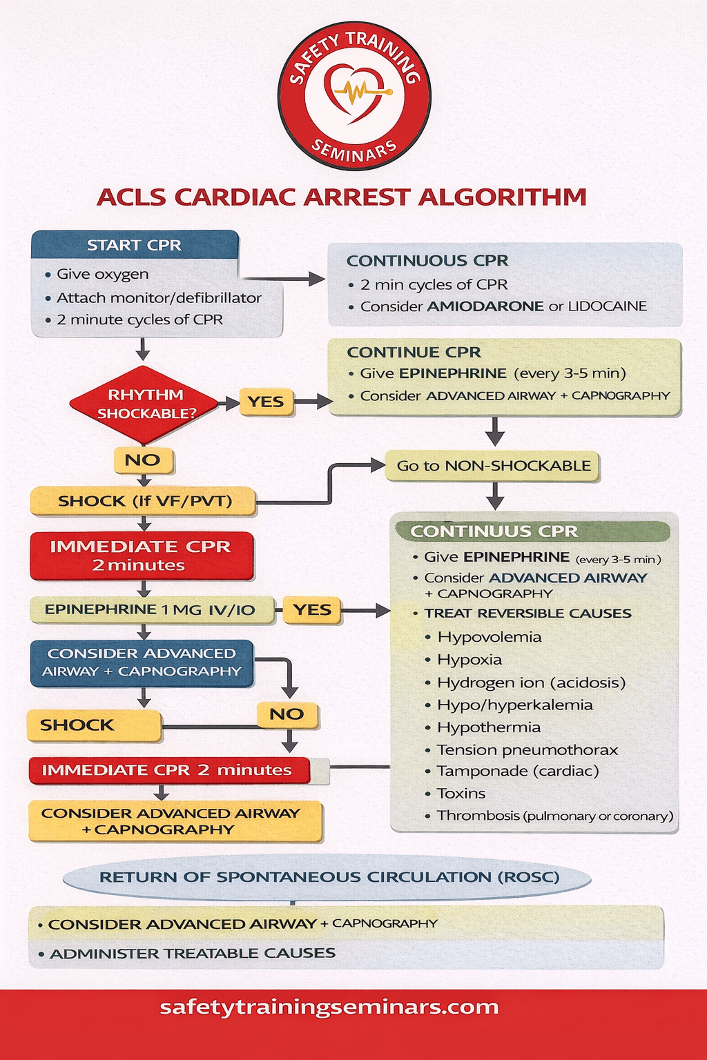
The ACLS Cardiac Arrest Algorithm provides a clear, step-by-step framework for managing adult cardiac arrest with precision and confidence. This infographic is designed to visually explain each critical action required during resuscitation, helping learners quickly grasp what to do, when to do it, and why it matters. It begins with rapid recognition of cardiac arrest, immediate activation of emergency response, and the initiation of high-quality CPR. The visual layout reinforces correct compression rate, depth, full chest recoil, and minimal pauses—core elements that directly impact patient survival.
As the algorithm progresses, the infographic highlights rhythm recognition as a key decision point. Providers are guided to distinguish between shockable rhythms, such as ventricular fibrillation and pulseless ventricular tachycardia, and non-shockable rhythms, including asystole and pulseless electrical activity. For shockable rhythms, the infographic emphasizes early defibrillation paired with continuous CPR. For non-shockable rhythms, it clearly outlines the importance of timely epinephrine administration and ongoing chest compressions. Medication timing, airway strategies, and the role of waveform capnography are visually mapped to improve understanding and retention.
The final section of the infographic focuses on continuous reassessment and post–cardiac arrest care. Every two minutes, providers are reminded to reassess rhythm and pulse while identifying and treating reversible causes using the Hs and Ts approach. Once ROSC is achieved, the algorithm shifts toward stabilizing oxygenation, ventilation, blood pressure, and neurological function. At Safety Training Seminars, this infographic-based explanation transforms the ACLS Cardiac Arrest Algorithm into an easy-to-follow, practical tool that supports confident decision-making during real-life cardiac emergencies.
The ACLS cardiac arrest algorithm guides healthcare providers in responding to cardiac arrest. It emphasizes high-quality CPR, early defibrillation, and advanced interventions. Safety Training Seminars teaches the latest AHA protocols for effective resuscitation.
High-quality CPR includes compressions at 100–120 per minute, 2–2.4 inch depth, full chest recoil, and minimal interruptions. Proper technique improves survival and is a core component of the ACLS cardiac arrest algorithm.
Defibrillation is applied immediately for shockable rhythms like ventricular fibrillation (VF) and pulseless ventricular tachycardia (pVT). Early shocks maximize return of spontaneous circulation. Safety Training Seminars teaches timely defibrillation steps.
Epinephrine is used every 3–5 minutes for non-shockable rhythms, and amiodarone or lidocaine for refractory VF/pVT. Medications combined with CPR and defibrillation improve patient outcomes.
Healthcare professionals including nurses, paramedics, and physicians benefit most. Training ensures readiness for real-life emergencies. Safety Training Seminars provides practical ACLS courses aligned with AHA guidelines.